Abstract
In children, hydroureteronephrosis due to ureterovesical stenosis may develop from reflux or obstruction. In diagnostic cystoscopy, not only the findings of the ureteral orifice and ureteral tracing but also the findings of accompanying intra- and extravesical pathologies should not be overlooked. A five-year-old male patient had increased left hydroureteronephrosis on ultrasound follow-up and Mag-3 scintigraphy showed partial obstruction. Retrograde pyelography (RPG) was planned for diagnostic purposes. Cystoscopy showed pulsation in the subureteric area of the left ureteral orifice. RPG was performed. Distal passage problems and proximal hydroureteronephrosis were found on the left side. Postoperative abdominal computed tomography (CT) angiography was performed to investigate the cause of arterial pulsation. It was observed that the left iliac artery did not bifurcate at the normal level and was aberrant in the pelvis as an external iliac artery, single arterial structure that was tortuous and slightly wide. At this level, it extended into the posterior neighborhood of the bladder. This anatomical variation and the suburethral neighborhood of the artery caused marked intravesical arterial pulsation. No intervention was performed, and it was noted that if surgical intervention was necessary according to the follow-up, it should be performed in absolute extravesical exploration, considering the existing pathology. Pulsatile mass detected during cystoscopy for intravesical evaluation may be associated with rare extravesical vascular malformations. Following CT angiography, invasive interventions should be carefully planned if necessary. Expansion of invasive procedures without a thorough understanding of extravesical anatomy may lead to serious complications.
Keywords: Hydroureteronephrosis, ureterovesical stenosis, iliac artery, vascular pathologies, hydronephrosis
Introduction
In children, a ureter diameter greater than 7 mm is considered megaureter (). Primary megaureter occurs due to a functional or anatomical abnormality of the ureterovesical junction. Primary megaureter is classified according to the presence or absence of reflux and obstruction ().
Primary obstructive megaureter accounts for approximately 25% of childhood obstructive uropathies (). It is frequently found in the left ureter and is bilateral in 10-15% of cases. Boys are affected more frequently than girls ().
Primary megaureter is often diagnosed by ultrasonography. Patients may present with symptoms such as urinary tract infection, abdominal pain, hematuria, or uremia. Some cases are diagnosed incidentally. Associated ipsilateral ureteropelvic junction obstruction, contralateral vesicoureteral reflux, and renal hypoplasia/dysplasia may be observed. Surgical intervention is required in only 10-30% of cases diagnosed with primary obstructive megaureter. It is believed that ureteral dilatation in patients with primary obstructive megaureter may improve over time in the natural course of the disease (-).
Case Presentation
A five-year-old male patient had been referred to the pediatric urology outpatient clinic at the age of three due to left hydronephrosis. The patient was asymptomatic during follow-up. The table below outlines the ultrasound follow-up conducted for the patient after reaching three years of age.
At the age of 3 years, voiding examination revealed no vesicoureteral reflux. Mag-3 scintigraphy indicated reduced and delayed perfusion and concentration function in the left kidney, along with a partial response to diuretics. The left kidney’s contribution to renal function was calculated at 51%.
During a follow-up Mag-3 scintigraphy at the age of 4.5 years, the patient exhibited increased hydroureteronephrosis on ultrasound. Activity retention was noted within dilated pelvicaliceal structures, followed by excretion post-diuretic injection. It was observed that the ureter became more prominent and tortuous during excretion. The left kidney’s contribution was recalculated at 48%.
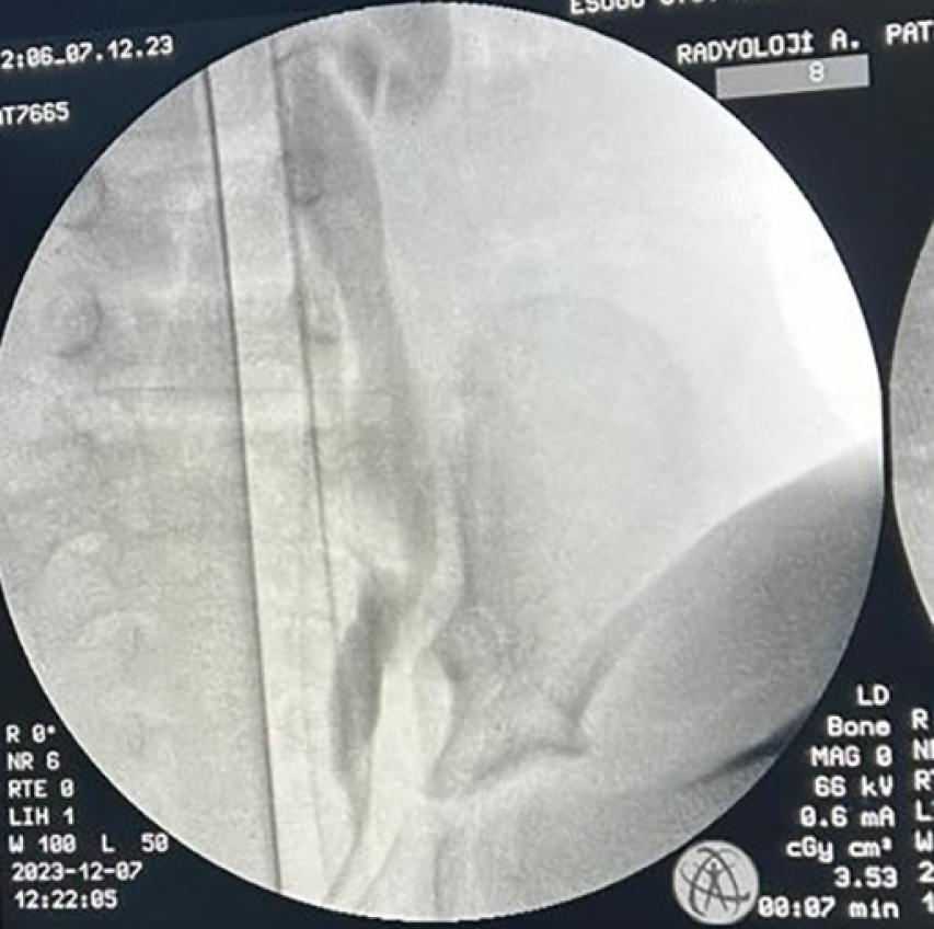
During the diagnostic cystoscopy of the patient, the ureters were observed to be in their normal anatomical location and structure. However, a pulsating formation protruding into the bladder was noticed superior to the left ureteral orifice. To address this, a 3 Fr ureteral stent was inserted into the left distal ureteral orifice, followed by retrograde pyelography (RPG). The RPG revealed mild stenosis of the ureteral orifice, characterized by significant tortuosity and a fold at the pelvic orifice ().
Due to the significant pulsation observed, computed tomography (CT) angiography was scheduled for further evaluation and treatment ().
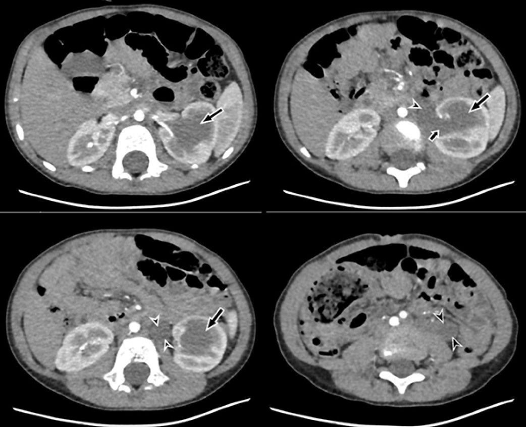
Abdominal CT angiography revealed grade 4 hydronephrosis in the left kidney, with the left ureter exhibiting tortuosity along its course. Notably, the abdominal aorta was positioned on the right side inferiorly to the origin of the renal arteries, with its bifurcation occurring at the level of the L3 vertebra. The left common iliac artery appeared narrow in caliber and anteriorly situated compared to the right, with the inferior mesenteric artery arising from it. The left common iliac artery did not bifurcate at the usual level; instead, it as a single artery resembling the left external iliac artery, displaying an aberrant course within the pelvis. The left internal iliac artery was observed to originate from this aberrant common iliac artery in the posterolateral vicinity of the inferior part of the bladder. Furthermore, the left external iliac artery exhibited indentation in the posterior vicinity of the bladder during its abnormal course. At this level, it appeared tortuous and slightly dilated (, , , , ) ().
CT: Computed tomography
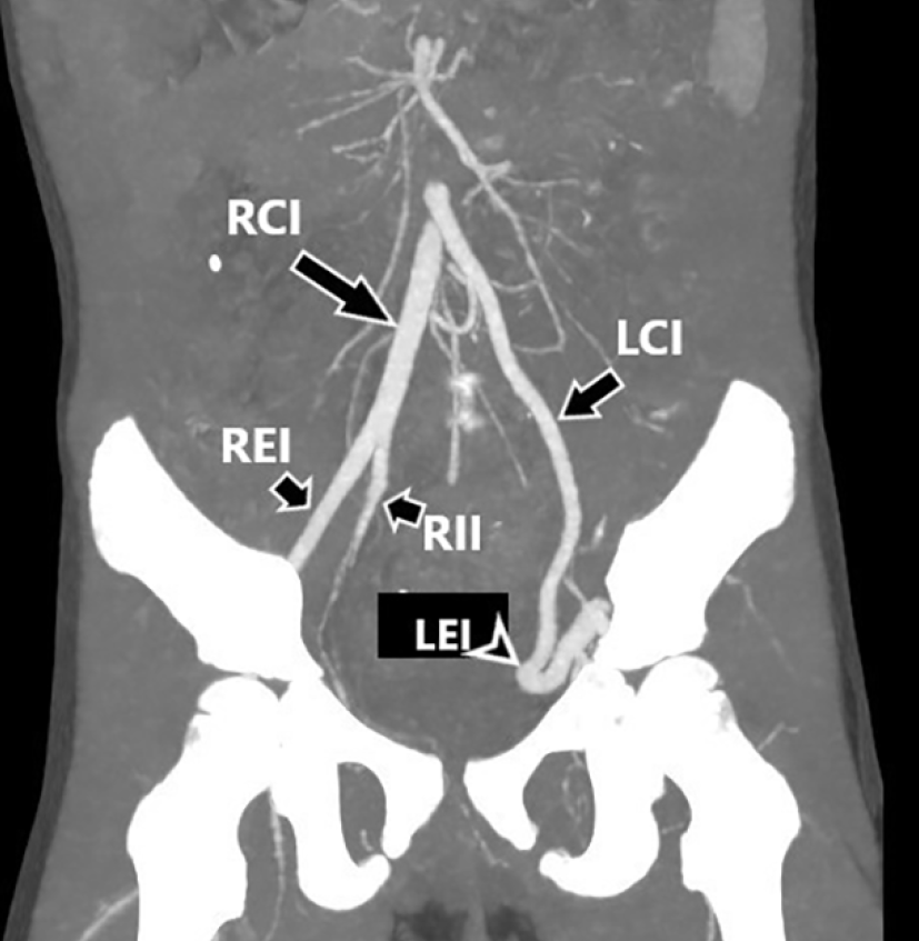
CT: Computed tomography, MIP: Maximum intensity projection, REI: Right external iliac, RII: Right internal iliac, RCI: Right common iliac, LCI: Left common iliac, LII: Left internal iliac, LEI: Left external iliac
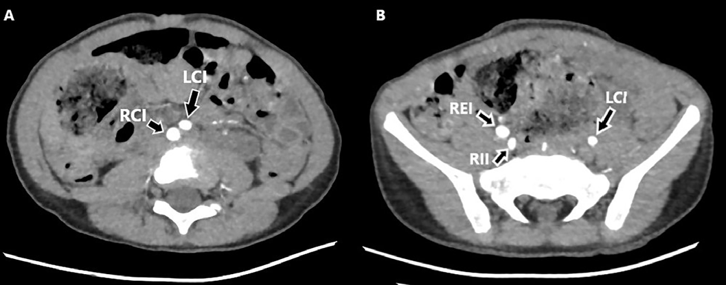
CT: Computed tomography, RCI: Right common iliac, LCI: Left common iliac, REI: Right external iliac, RII: Right internal iliac
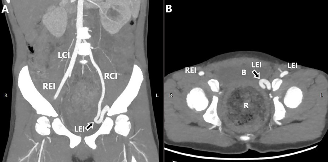
MIP CT angiography images show normal RCI and REI. The LCI does not bifurcate at a normal level, but continues distally into the pelvis as a single artery. The LCI branches off from the LII in the pelvis, and continues as a tortuous and dilated LEI artery. The LEI shows an indentation to the left posterolateral wall
B: Bladder, R: Rectum, REI: Right external iliac, RCI: Right common iliac, LCI: Left common iliac, LII: Left internal iliac, LEI: Left external iliac, MIP: Maximum intensity projection
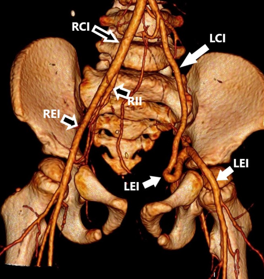
CT: Computed tomography, REI: Right external iliac, RII: Right internal iliac, RCI: Right common iliac, LCI: Left common iliac, LII: Left internal iliac, LEI: Left external iliac
Table 1. Follow-up of the patient’s urinary system ultrasounds
- | 3 years old | 4 years old | 4 years 6 months old | |
|---|---|---|---|---|
Kidney dimension | Right | 61x25 mm | 67x37 mm | 69x42 mm |
Left | 77x38 mm | 78x40 mm | 96x40 mm | |
Anterior-posterior diameter | Right | - | - | - |
Left | 13 mm | 15 mm | 19 mm | |
Degree of hydronephrosis | Right | - | - | - |
Left | Grade 3 | Grade 3-4 | Grade 3-4 | |
Parenchyma | Right | 6 mm | 9 mm | 11 mm |
Left | 6 mm | 7 mm | 9 mm | |
Ureteral dilatation | Right | - | - | - |
Left | 11 mm | 15 mm | 15 mm | |
Discussion
Primary obstructive megaureter is a rare urinary system anomaly. The pathogenesis of primary obstructive megaureter has not been fully elucidated. It is attributed to delayed or abnormal muscle development in the distal ureter, which should occur in the first week of gestation (). This leads to the formation of an aperistaltic segment causing functional obstruction. Rarely, megaureter occurs due to congenital ureteral stricture or valves (, ). The prognosis is generally good for unilateral ureterovesical strictures diagnosed antenatally. There are no randomized studies indicating the optimal treatment for patients with primary obstructive megaureter. In many cases, hydroureteronephrosis regresses spontaneously (, ). Spontaneous recovery occurs with segmental maturation. The spontaneous resolution time is prolonged in cases with severe hydroureteronephrosis. The chance of spontaneous resolution is lower in cases with scintigraphically demonstrated obstructive lesions. If there is no additional congenital renal abnormality, long-term results are favorable. Surgical treatment is indicated if hydronephrosis increases or renal function decreases during follow-up. Recurrent urinary tract infections, pyelonephritis, persistent flank pain, or hematuria also require surgical intervention. Ten percent of cases with megaureter undergo surgery due to decreased renal function at the initial evaluation.
While complete resolution is observed in 34% of cases during conservative follow-up, 49% of cases remain stable (). In some cases, increased dilatation, urinary tract infection, and decreased renal function necessitate surgical intervention within the first year. Reimplantation of the dilated ureter in small infants is technically challenging and can potentially lead to bladder dysfunction (). Therefore, less invasive procedures such as endoscopic or open Double-J catheter placement are preferred (, ). This approach helps to alleviate the obstruction and reduces the need for reimplantation (, ). In the literature, endoscopic catheter placement has been successful in approximately 30% of cases (). In our patient, we initially aimed to perform an examination for etiological investigation without placing a catheter. We believed that interventions could result in serious complications.
Conclusion
The pulsatile mass identified during cystoscopy in the intravesical assessment may be linked to significant extravesical vascular malformations. Invasive interventions should be carefully planned following CT angiography. Extending invasive procedures without a comprehensive understanding of the extravesical anatomy could lead to serious complications. Therefore, a thorough evaluation of the vascular anatomy is imperative to ensure safe and effective management of the patient’s condition.
After diagnostic evaluation, open or laparoscopic extravesical exploration, and ureteral surgery can be planned if necessary, according to the findings.
Footnotes
Ethics
Informed Consent: Written informed consent was obtained from the patient.
Authorship Contributions
Surgical and Medical Practices: D.D., B.T., Concept: D.D., B.T., Design: D.D., E.Ş., Data Collection or Processing: D.D., Ç.Ö., Analysis or Interpretation: D.D., B.T., Literature Search: D.D., Writing: D.D.
Conflict of Interest: No conflict of interest was declared by the authors.
Financial Disclosure: The authors declare that they received no financial support for this study.
References
Cussen LJ. Dimensions of the normal ureter in infancy and childhood. Invest Urol. 1967;5:164-178.
King LR. Megaloureter: definition, diagnosis and management. J Urol. 1980;123:222-223.
Liu HY, Dhillon HK, Yeung CK, Diamond DA, Duffy PG, Ransley PG. Clinical outcome and management of prenatally diagnosed primary megaureters. J Urol. 1994;152:614-617.
Shukla AR, Cooper J, Patel RP, Carr MC, Canning DA, Zderic SA, Snyder HM 3rd. Prenatally detected primary megaureter: a role for extended followup. J Urol. 2005;173:1353-1356.
McLellan DL, Retik AB, Bauer SB, Diamond DA, Atala A, Mandell J, Lebowitz RL, Borer JG, Peters CA. Rate and predictors of spontaneous resolution of prenatally diagnosed primary nonrefluxing megaureter. J Urol. 2002;168:2177-2180.
Gimpel C, Masioniene L, Djakovic N, Schenk JP, Haberkorn U, Tönshoff B, Schaefer F. Complications and long-term outcome of primary obstructive megaureter in childhood. Pediatr Nephrol. 2010;25:1679-1686.
Calisti A, Oriolo L, Perrotta ML, Spagnol L, Fabbri R. The fate of prenatally diagnosed primary nonrefluxing megaureter: do we have reliable predictors for spontaneous resolution? Urology. 2008;72:309-312.
Tanagho EA. Intrauterine fetal ureteral obstruction. J Urol. 1973;109:196-203.
Cuchi JA, Chandran H. Congenital ureteral strictures; a rare cause of antenatally detected hydronephrosis. Pediatr Surg Int. 2005;21:566-568.
Kannaiyan L, Karl S, Mathai J, Chacko J, Sen S. Congenital ureteric stenosis: a study of 17 children. Pediatr Surg Int. 2009;25:513-517.
Lipski BA, Mitchell ME, Burns MW. Voiding dysfunction after bilateral extravesical ureteral reimplantation. J Urol. 1998;159:1019-1021.
Farrugia MK, Steinbrecher HA, Malone PS. Use of stents in the management of primary obstructive megaureters requiring intervention before the age of one year. J Pediatr Urol. 2011;7:198-202.
Shenoy MU, Rance CH. Is there a place for JJ stent placement as a temporary procedure for symptomatic partial congenital vesico-ureteric junction obstruction in infancy? BJU Int. 1999;84:524-525.
Barbancho DC, Fraile AG, Sanchez RT, Diaz ML, Fernandez MM, Vazquez FL, Bramtot AA. Is initial treatment of primary nonrefluxed megaureter with double-J stent effective? Cir Pediatr. 2008;21:32-36.
Supplementary Materials
Video 1
Download