Abstract
This study systematically reviewed how boron exposure affects animal model sperm parameters and testicular structure. The Embase, Scopus, PubMed, ProQuest, and Web of Science databases were searched up to January 2023. The studies that examined boron’s efficacy and safety regarding semen parameters and testicular histopathology in different animal species were included. Studies involving specific derivatives or in vitro or human studies were excluded. Two reviewers selected the studies and extracted the data independently. The quality of eligible studies was assessed using the Animal Research: Reporting of In Vivo Experiments Essential 10 checklist. The outcomes were summarized and presented in tables. Sixteen studies were included from 1,602 retrieved articles. While some studies demonstrated that boron, at doses of 17.5, 35, and 70 mg/kg for 8 weeks, improved the quality of spermatogenesis in terms of rate of sperm movement, sperm concentration, and total volume of sperm, other studies showed dose-dependent boron toxicity to the reproductive system. While some studies suggest potential benefits of boron supplementation on spermatogenesis, others indicate harmful effects. The conflicting results emphasize the need for further research to establish clear guidelines on the appropriate dosage, duration, and safety of boron in improving sperm quality.
Keywords: Boron, sperm parameters, testicular histopathology, reproductive toxicity, systematic review
Introduction
Boric acid, containing boron, is an inorganic acid often used as a water pH regulator, stabilizer, neutralizer, and buffer in the glass industry, food preparation, beauty industry, agriculture, and pharmaceutical production (, ). Obviously, with the development of the industry, the importance of this acid and its amount in our environment has increased due to its widespread use. In recent years, the physiological role of boron has attracted the attention of researchers worldwide, especially in the field of the reproductive system (). Studies on the effects of boron on animals began in the 1990s. According to the European Union report, boron is placed in group 1B of the Globally Harmonized System classification, specifically in the category R60-61. Group R60 weakens the reproductive system, while group R61 is harmful to the fetus ().
Although the definite role of boron in the physiology of animals is not completely clear, the effects of its supplementation in the diet on bone growth and in the central nervous, endocrine, and reproductive systems have been reported (, ). Boron increases the level of interleukin-6 and regulates serine protease enzyme activity. It also improves the immune and the antioxidant capacity and calcium metabolism (). In animals, most of the studies on the effects of boron focus on mice and rats. The findings of these studies are interesting because they have produced contradictory results regarding Boron’s activity. The study showed that high concentrations of boron have positive effects on lipid peroxidation reduction and antioxidant metabolism in mice (). In rabbits, boron improves antioxidant capacity, sperm quality, and testosterone concentration (). However, high concentrations of boron have negative effects on the reproductive system of animals. Boric acid causes testicular atrophy in rats and dogs at specific doses and concentrations (). Another study found that a diet with 1,000 ppm boron, reduced the number of spermatocytes, spermatids, and mature spermatozoa in mice after 30 days (). Additionally, high dietary boron concentrations have detrimental effects on the reproductive system of male rats, and at a certain dose, these concentrations can lead to acute toxicity and death. In animal tissue studies, toxic concentrations of boron damage the process of spermatogenesis, resulting in testicular atrophy in 10-14 days ().
According to studies on the effect of boron on the reproductive system of animals, it is believed that boron acts as a double-edged sword that can have beneficial or harmful effects on this system, depending on the dose. Therefore, in this study, we aimed to systematically review the effect of boron on sperm parameters and testicular histopathology in animal models.
Methods
This systematic review followed the guidelines of the preferred reporting items for systematic reviews and meta-analyses (PRISMA) Statement and the Cochrane Collaboration Handbook. Due to resource limitations and institutional policies at the time, registration was not feasible; furthermore, the narrowly defined scope of this review was considered sufficient to justify the absence of registration.
Data Sources and Search Strategy
The Embase, Scopus, PubMed, ProQuest, and Web of Science databases were thoroughly searched in January 2023. Finding both published and unpublished studies was the goal of the search approach. A three-step search process was used in this study. Following a preliminary, constrained search of MEDLINE, the title and abstract’s text words were examined. In January 2023, a second search across all included databases was conducted using all indicated keywords and index phrases: ((((“reproduction”[MeSH Terms]) OR (“reproduction”[Title/Abstract])) OR (“reproductive”[Title/Abstract])) OR (((“infertility”[MeSH Terms]) OR (infertil*[Title/Abstract])) OR (Sperm [Title/Abstract]))) AND ((“boron”[MeSH Terms]) OR (boron [Title/Abstract])).
The last step was to look for further studies in the reference lists of all the indicated papers and articles. This review covered studies that were released on any date and in any language.
Inclusion and Exclusion Criteria
The original studies that examined boron’s efficacy and safety of semen parameters and testicular histopathology in different animal species were included. Studies involving specific derivatives or in vitro/human studies were excluded. Also, Abstracts, reviews, letters, and theses were excluded.
Study Selection and Data Extraction
The retrieved articles from multiple information sources were organized using PRISMA flowcharts. Two reviewers independently screened all titles and abstracts of the retrieved articles. Additionally, full texts of relevant studies were independently assessed for eligibility, with reasons for exclusion documented for the full texts that were excluded. Data extraction was performed separately by two researchers, and any discrepancies were discussed and resolved. The following data was extracted from the included studies: Author Name, Publishing Year, Country, Study Design, Animal, Sample Size, Age, Diet, Boron Derivative and Dosage, Intervention Duration, Testis Weight, Testicular Morphometry, Pathology Report, Sperm Count/Concentration, Sperm Abnormality, Sperm Motility, and Ejaculate Volume.
Risk of Bias Assessment
Using the Animal Research: Reporting of In Vivo Experiments (ARRIVE) Essential 10 checklist, two independent reviewers evaluated the methodological quality of the selected articles. The reviewers’ probable differences were settled through conversation or by a third reviewer.
Data Synthesis
The primary outcomes in this study were the change of sperm parameters and testis histopathology in animals with boron exposure. As these outcomes were diverse or heterogeneous (from different animal species), combining the data and conducting a meta-analysis was not possible. The results are summarized and presented in tables.
Results
Study Selection
The electronic search, manual search, and reference check yielded a total of 1,602 citations. After removing duplicate citations, we were left with 919 studies for screening. Based on the titles and abstracts provided, 35 papers were selected, while 19 articles were disqualified during the full-text selection process. Ultimately, 16 studies were included. For more detailed information on the selection procedure, please refer to in the PRISMA flowchart.
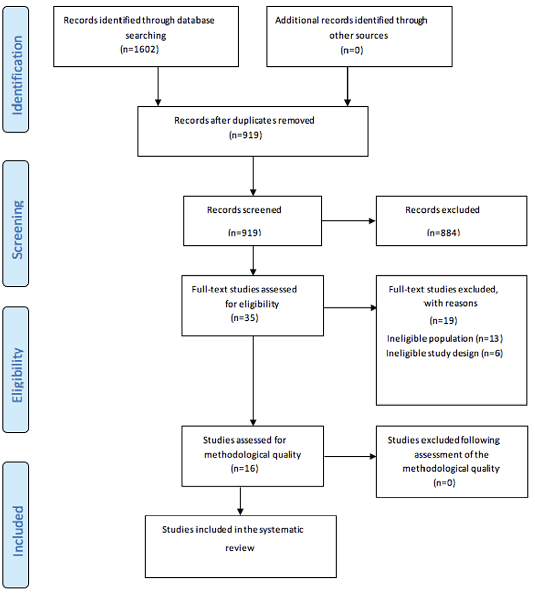
PRISMA: Preferred reporting items for systematic reviews and meta-analyses
Study Characteristics
In this research, 16 studies were reviewed from 1976 to 2022. These studies were conducted in the United States, Japan, Chile, Egypt, Türkiye and India. The investigated animals were rats, mice, rabbits, African clawed frogs, goats, and Osemi Rams.
Results of Individual Studies
As mentioned, the results of the studies were notably different from each other. In the study of Elkomy et al. (), Ibrahim et al. (), Krishnan et al. (), and Abdel-Wahab et al. (), boron improved the quality of spermatogenesis. In the Elkomy et al. () study, after the administration of boron at the doses of 17.5, 35, and 70 mg/kg for 8 weeks, it was seen that the rate of sperm movement, sperm concentration, and total volume of sperm increased compared to the control group (). Similarly, the study by Ibrahim et al. (), which gave a 70 mg/kg boron-containing diet to Osmei rams for 4 months, showed an improvement in total sperm volume and sperm concentration (). Krishnan et al. () also administered 40 ppm of this acid in the form of sodium tetraborate to goats for 60 days and concluded that there was a significant increase in sperm motility compared to the control group. Another interesting result was obtained by Abdel-Wahab et al. () in 2022 by administering 70 mg/kg of boric acid to goats for 24 weeks; they observed that goats receiving boron exhibited improvements in the structure of testis tubules, and the activity of Sertoli cells increased. Conversely, other studies have shown that boron toxicity affects the reproductive system. Ayranci et al. () administered 1000 mg/kg/day of boric acid to Sprague-Dawley rats for 7 days, and observed an increase in edema in the testicular interstitial tissue and an increase in apoptotic cells. El-Dakdoky et al. () also gave 125, 250, or 500 mg/kg of boron to Wistar rats for 60 days, which led to the destruction of spermatogonia, spermatocytes, and spermatozoa with a dose of 250 mg/kg and severe destruction of germ cells with a dose of 500 mg/kg. Lee et al. () administered boron to Sprague-Dawley rats with doses of 500-1000-2000 ppm for 90 days. They observed that with a dose of 500 ppm, boron did not cause any complications. However, with a dose of 1000 ppm, germ cell destruction began, and the diameter of seminiferous tubules was reduced. Eventually, testicular atrophy occurred. and represent the characteristics of the included studies.
Table 1. Baseline characteristics of studies
No | Author (year) | Country | Study design | Animal | Population size | Age | Diet | Boron | Treatment duration | |||
|---|---|---|---|---|---|---|---|---|---|---|---|---|
Boron | Control | Derivative | Dosage | Dosage in control | ||||||||
1 | Dixon et al. () (1976) | USA | Serial mating study | Sprague-Dawley rats | 30 | - | - | - | Borax (11.3% boron) | Three groups: 0.3, 1.0, or 6.0 mg/L of drinking water | - | 90 days |
2 | Lee et al. () (1978) | USA | Randomized controlled trial | Sprague-Dawley rats | 54 | 18 | - | - | Borax | 500, 1000, and 2000 ppm | 0 | 60 days |
3 | Treinen and Chapin () (1991) | USA | Randomized controlled trial | Fischer 344 (CDF (F344)/ CrlBr) rats | 36 | 30 | 120 day | Powdered NIH-07 feed | Boric acid | 9000 ppm | 0 | 4 weeks |
4 | Ku et al. () (1993) | USA | Randomized controlled trial | Fischer 344 (CDF (F344) rats | 216 | 54 | 60 to 70 days | Powdered NIH-07 | Boric acid | 3000, 4500, 6000, or 9000 ppm | B levels <20 ppm | 9 weeks |
5 | Chapin and Ku (1994) () | USA | Reproductive assessment by continuous breeding study | CD-1 mice | 6 | 4 | 11 weeks | - | Boric acid | 1000, 4500, and 9000 ppm | - | 28 days |
6 | Nomiyama et al. () (1996) | Japan | Randomized controlled trial | Wister rat | 24 | 12 | 13 weeks | Pellet and distilled water ad libitum | Diborane via inhalation | Two groups: 0.1 or 1.0 ppm for 6 hr/day, 5 days/week | 0 | 8 weeks |
7 | Bustos-Obregon et al. () (2007) | Chile | Randomized controlled trial | Mice (Mus musculus, CF-1 strain) | 10 | 10 | 85 days | Commercial pellet and water ad libitum | - | 12 mg of boron/L in drinking water | 0.6 mg boron/L in drinking water | 42 days |
8 | Espinoza-Navarro et al. () (2007) | Chile | Randomized controlled trial | Mice (CF-1 strain) | 32 | 16 | - | Standard conditions of living | Boric acid | Two groups: 2.0 to 6.0 mg boron/L in drinking water and 12.0 mg boron/L in drinking water | 0.54 mg boron/L in drinking water | 60 days |
9 | Bustos-Obregón and Olivares () (2012) | Chile | Randomized controlled trial | mice (Mus domesticus, CF-1 strain) | 10 | 10 | 85 days | Standard animal room conditions | - | 12 mg boron/L in drinking water | 0.6 mg boron/L in drinking water | 42 days |
10 | El-Dakdoky and Abd El-Wahab (16) (2013) | Egypt | Randomized controlled trial | Wister rat | 24 | 8 | 12 weeks | Standard laboratory pellets and water ad libitum | Boric acid | Three groups: 125-250-500 mg boric acid/kg body weight/day | 0 | 60 days |
11 | Elkomy et al. () (2015) | Egypt | Randomized controlled trial | V. line rabbits | 15 | 5 | 10 months | Pellet diet ad libitum | Boric acid | Three groups: 17.5, 35, 70 mg boron /kg | 0 | 8 weeks |
12 | Fort et al. () (2016) | USA | Randomized controlled trial | African clawed frog xenopus laevis | 48 | 12 | 3 years | Salmon starter - dry pellets ad libitum | Boric acid | Four groups: 5 - 7.5 -10 - 15 mg boron/L equivalent to 28.5, 42.8, 57.0, and 85.5 mg boric acid/L | 0 | 30 days |
13 | Ibrahim et al. () (2019) | Egypt | Randomized controlled trial | Osemi rats | 6 | 6 | 4 months | Basal ration ad libitum | Boric acid | 400 mg boric acid/kg diet=70 mg boron/kg diet | 0 | 4 months |
14 | Krishnan et al. () (2019) | India | Randomized controlled trial | Goat | 7 negative control, 7 selenium supplement as positive control | 7 | - | 55% roughage and 45% concentrate | Sodium tetra borate | 40 ppm in diet | 0 | 60 days |
15 | Ayranci et al. () (2021) | Turkiye | Randomized controlled trial | Sprague-Dawley type albino rats | 30 | 12 | 12 weeks | - | Boric acid | 1000 mg/kg/day added to the drinking water | 0 | 7 days |
16 | Abdel-Wahab et al. () (2022) | Egypt | Randomized controlled trial | Goat | 6 | 6 | 4 months | Normal basal composition, roughage and berseem ad libitum | Boric acid | 400 mg boric acid/kg diet=70 mg boron/kg diet | 0 | 24 weeks |
Table 2. Summary of the results
No. | Author (year) | Sample type | Testis weight | Testicular morphometry | Sperm count/conc. | Sperm abnormality | Sperm motility | Ejaculate volume | Pathology report |
|---|---|---|---|---|---|---|---|---|---|
1 | Dixon et al. () (1976) | Testicular tissue | (g) 0.3 boron: 1.64±0.67, 1 boron:1.66±0.49, 6 boron: 1.71±0.10 | - | - | - | - | - | Without significant reproductive toxicity. |
2 | Lee et al. () (1978) | Testicular tissue | (g) control: 1.81±0.06, 500 ppm boron: 1.76±0.19, 1000 ppm boron: 0.68±0.16*, 2000 ppm boron: 0.63±0.01* | - | - | - | - | - | At 500 ppm, there were no significant adverse effects observed. In contrast, male rats receiving 1000 and 2000 ppm of boron displayed a significant loss of germinal elements, decrease in seminiferous tubular diameter, and accumulation of testicular boron. The testicular atrophy was greatest at the highest dose, and depletion of germ cells was complete after 60 days of exposure. |
3 | Treinen and Chapin () (1991) | Testicular tissue | - | - | - | - | - | - | First testicular lesion noted was an inhibition of spermiation, which appeared on day 7. Widespread exfoliation of apparently viable germ cells and pachytene cell death in stages VII and XIV appeared as exposure continued. After 28 days of dosing, extreme epithelial disorganization and [effect] were observed, indicating the need for further investigation. germ cell loss were evident. |
4 | Ku et al. () (1993) | Testicular tissue | - | - | - | - | - | - | Inhibited spermiation was most reliably reflected by detailed testicular histology, with the more severe cases decreasing epididymal sperm count to levels that could affect fertility. |
5 | Chapin and Ku (1994) () | Testicular tissue | (mg) Control: 140±3, 1000 ppm: 140±4, 4500 ppm: 69±5*, 9000 ppm: 20±1* | - | Concentration control: 518.6±35.8, 1000 ppm: 532.4±40.9, 4500 ppm: 146.9±26.6 *, 9000 ppm: 2.8±1.7 * | - | % control: 78.1±3.0, 1000 ppm: 69.0±4.5*, 4500 ppm: 53.3±8.2*, 9000 ppm: 42.9 | - | The first lesion appeared in some animals at day 7, and consisted of an inhibition of sperm release. This progressed in severity, and was soon (day 21) accompanied by a disorganization of the epithelium and the release of immature germ cells. By day 28, there were some atrophic tubules that contained only residual spermatogonia and the somatic Sertoli cells. |
6 | Nomiyama et al. () (1996) | Testicular tissue | - | - | Count (106) in head of epididymis: control: 69.1±18.8, 0.1 ppm: 76.5±21.3, 1 ppm: 76.5±9.2 count in body/tail of epididymis: control: 262.5±55.2, 0.1 ppm: 244.1±32.5, 1 ppm: 267.7±35.5 | Head abnormality (103) abnormal type: control: 12.8±4.2, 0.1 ppm: 14.3±5.1, 1 ppm: 10.2±6.2 immature type: control: 11.3±4.0, 0.1 ppm: 12.8±4.7, 1 ppm: 9.1±5.9 | - | - | There were no differences between the testes of control and exposed rats. |
7 | Bustos-Obregón et al. () (2007) | Testicular tissue | - | (μm) Tubular diameter*: control: 208.9±5.76, boron: 233.3±9.91 epithelial height*: control: 50.4±0.88, boron: 58.3±3.01 tubular lumen*: control: 60.5±1.41, boron: 72.6±4.10 | - | - | - | - | The boron-exposed group exhibits a 27% of histological changes, with a 2% of tubular atrophy, a 7% of tubular obstruction and an 18% of epithelial vacuolization. In the control group, damage was less than 1%. |
8 | Espinoza-Navarro et al. () (2007) | Testicular tissue | (g) control: 0.8±0.834, 2.0-6.0 mg boron: 1.1±0.106*, 12.0 mg boron: 1.3±0.075* | - | - | - | - | - | In boron groups the basal epithelium of seminal tubules of the testicles were altered, with an increase in the lumen and an absence of spermatozoids and great presence of vacuolization in the germinative epithelium. |
9 | Bustos-Obregón and Olivares () (2012) | Testicular tissue | - | (μm) Tubular diameter*: control: 189±2.25, boron: 155±3.47 luminal diameter*: control: 60.2±1.16, boron: 78.1±1.24 epithelial height*: control: 61.0±1.12, boron: 50.7±0.89 tunica albuginea thickness*: control: 107.6±28.4, boron: 29.70±9.1 interstitial area* (% area): control: 14.4±1.00, boron: 29.2±6.57 | - | - | - | - | Boron produces vacuolization, tubular epithelial desquamation and tamponade. |
10 | El-Dakdoky and Abd El-Wahab () (2013) | Testicular tissue | Right testis (g): control: 1.57±0.03, 125 mg boric acid/kg/day: 1.49±0.04, 250 mg: 1.38±0.10*, 500 mg: 0.46±0.02* | Seminiferous tubules diameter (μm): control: 234.6±2.48, 125 mg B: 231.7±4.65, 250 mg B: 219.2±2.61*, 500 mg B: 143.0±1.14* germinal cell thickness (μm): control: 70.10±1.11, 125 mg B: 67.34±1.85, 250 mg B: 60.18±0.75*, 500 mg B: 21.00±0.60* | Count (106): control: 36.47±3.94, 125 mg B: 33.70±2.69, 250 mg B: 16.8±1.73*, 500 mg B: 0.03±0.02* | (%) control: 3.10±0.2, 125 mg B: 4.10±0.51, 250 mg B: 12.36±1.58* | - | - | Moderate testicular degeneration in B-250 group, the numbers of spermatogonia, spermatocytes and spermatozoa were diminished, vacuolation and degenerative cells in the basal region of the tubules were observed. In B-500 group, almost all germ cells disappeared from the atrophied STs and the testicular vessels appeared severely congested. |
11 | Elkomy et al. () (2015) | Semen analysis | - | - | Concentration (106): control: 228.56±4.18, 10 ppm: 259.00±2.95*, 200 ppm: 258.50±2.88*, 400 ppm: 265.38±2.83* count (106): control: 118.18±13.82, 10 ppm: 171.95±15.39*, 200 ppm: 161.03±13.37*, 400 ppm: 165.50±12.14* | (%) control: 10.34±1.00, 10 ppm: 4.08±0.46 *, 200 ppm: 4.85±0.53 *, 400 ppm: 3.98±0.35* | (%) control: 60.00±4.23, 10 ppm: 81.88±1.64, 200 ppm: 87.19±1.44*, 400 ppm: 91.88±1.01* | (mL) control: 0.51±0.06, 10 ppm: 0.66±0.06, 200 ppm: 0.62±0.05, 400 ppm: 0.62±0.03 | |
12 | Fort et al. () (2016) | Testicular tissue | - | - | Count (106): control: 2.2±0.03, 5 mg boron/L: 2.1±0.02, 7.5 mg boron/L: 2.1±0.03, 10.0 mg boron/L: 2.0±0.0, 15.0 mg boron/L: 1.8±0.1* | (%) control: 0.02±0.001, 5.0 mg boron/L: 0.02±0.001, 7.5 mg boron/L: 0.02±0.001, 10.0 mg boron/L: 0.03±0.003, 15.0 mg boron/L: 0.04±0.005* | - | - | The general appearance and location of the testes was normal and histological evaluation of the testes was not deemed necessary. |
13 | Ibrahim et al. () (2019) | Semen analysis | - | - | Sperm concentration/mm3: control: 1.35±0.06, boron: 3.35±0.07* | Primary (%): control: 2.00±0.37, boron: 1.67±0.33 secondary (%): control: 15.6±0.84, boron: 7.83±1.11* | - | (mL): control: 0.57±0.07, boron: 1.12±0.33* | - |
14 | Krishnan et al. () (2019) | Testicular tissue, Semen analysis | (g): control: 169.00 ±14.6, boron: 185.3±11.80 | - | - | - | Significantly higher total sperm motility (%) and total progressive motility (%) as compared to control | - | - |
15 | Ayranci et al. () (2021) | Testicular tissue | (g): control: 3.033±0.362, boron: 2.426±0.623* | - | - | - | - | - | Edema in the interstitial area in the testis tissue, increase in Leydig cells, basal membrane thickening in the seminiferous tubules, more apoptotic cells. |
16 | Abdel-Wahab et al. () (2022) | Testicular tissue | - | Diameter of seminiferous tubules: large tubules: control: 672.24±5.44, boron: 750.05±5.21*, small tubules: control: 510.04±3.66, boron: 548.06±3.55* height of spermatogenic cells: control: 154±1.21, boron: 180.22±1.19* | - | - | - | - | Boron supplementation succeeded remarkably in improving the testicular architecture and appeared with normal morphology and included seminiferous tubules lined by normal spermatogenic cells and Sertoli cells. The lumen of the tubules was noticed to include huge amount of spermatid and sperms. Also, the interstitial tissues were found to contain blood capillaries and interstitial cells. |
*: p-value<0.05
Red: Negative effect
Green: Positive effect
White: No difference
Methodological Quality
The risk of bias assessment of included studies is summarized in . All studies were of good general quality based on the ARRIVE Essential 10 appraisal checklist.
Table 3. Critical appraisal results of eligible studies
No | Study | Q1A | Q1B | Q2A | Q2B | Q3A | Q3B | Q4 | Q5 | Q6 | Q7A | Q7B | Q8A | Q8B | Q8C | Q9A | Q9B | Q10A | Q10B |
|---|---|---|---|---|---|---|---|---|---|---|---|---|---|---|---|---|---|---|---|
1 | Dixon et al. () (1976) | Y | Y | Y | N | Y | Y | Y | N | Y | Y | NA | Y | Y | Y | Y | N | Y | NA |
2 | Lee et al. () (1978) | Y | Y | Y | N | Y | Y | N | N | Y | Y | NA | Y | Y | Y | Y | N | Y | NA |
3 | Treinen and Chapin () (1991) | Y | Y | Y | N | Y | Y | Y | N | Y | Y | NA | Y | Y | Y | Y | Y | Y | NA |
4 | Ku et al. () (1993) | Y | Y | Y | N | Y | Y | Y | N | Y | Y | NA | Y | Y | Y | Y | Y | Y | NA |
5 | Chapin and Ku (1994) () | Y | Y | Y | N | Y | Y | N | N | Y | Y | NA | Y | Y | Y | Y | Y | Y | NA |
6 | Nomiyama et al. () (1996) | Y | Y | Y | N | Y | Y | N | N | Y | Y | NA | Y | Y | Y | Y | Y | Y | NA |
7 | Bustos-Obregón et al. () (2007) | Y | Y | Y | N | Y | Y | Y | N | Y | Y | NA | Y | Y | Y | Y | Y | Y | NA |
8 | Espinoza-Navarro et al. () (2007) | Y | Y | Y | N | Y | Y | N | N | Y | Y | NA | Y | Y | Y | Y | Y | Y | NA |
9 | Bustos-Obregón and Olivares () (2012) | Y | Y | Y | N | Y | Y | N | N | Y | Y | NA | Y | Y | Y | Y | N | Y | NA |
10 | El-Dakdoky and Abd El-Wahab () (2013) | Y | Y | Y | N | N | Y | N | N | Y | Y | NA | Y | Y | Y | Y | Y | Y | NA |
11 | Elkomy et al. () (2015) | Y | Y | Y | N | Y | Y | Y | N | Y | Y | NA | Y | Y | Y | Y | N | Y | NA |
12 | Fort et al. () (2016) | Y | Y | Y | N | Y | Y | Y | N | Y | Y | NA | Y | Y | Y | Y | N | Y | NA |
13 | Ibrahim et al. () (2019) | Y | Y | Y | N | Y | Y | N | N | Y | Y | NA | Y | Y | Y | Y | N | Y | NA |
14 | Krishnan et al. () (2019) | Y | Y | Y | N | Y | Y | N | N | Y | Y | NA | Y | Y | Y | Y | N | Y | NA |
15 | Ayranci et al. () (2021) | Y | Y | Y | N | Y | Y | Y | N | Y | Y | NA | Y | Y | Y | Y | N | Y | NA |
16 | Abdel-Wahab et al. () (2022) | Y | Y | Y | N | Y | Y | Y | N | Y | Y | NA | Y | Y | Y | Y | N | Y | NA |
Y: Yes, N: No, U: Unclear, NA: Not applicable; the ARRIVE Essential 10: Compliance Questionnaire: Q1A: Are all experimental and control groups clearly identified?; Q1B: Is the experimental unit (e.g. an animal, litter or cage of animals) clearly identified?; Q2A: Is the exact number of experimental units in each group at the start of the study provided (e.g. in the format ‘n=’)?; Q2B: Is the method by which the sample size was chosen explained?; Q3A: Are the criteria used for including and excluding animals, experimental units, or data points provided?; Q3B: Are any exclusions of animals, experimental units, or data points reported, or is there a statement indicating that there were no exclusions?; Q4: Is the method by which experimental units were allocated to control and treatment groups described?; Q5: Is it clear whether researchers were aware of, or blinded to, the group allocation at any stage of the experiment or data analysis?; Q6: For all experimental outcomes presented, are details provided of exactly what parameter was measured?; Q7A: Is the statistical approach used to analyse each outcome detailed?; Q7B: Is there a description of any methods used to assess whether data met statistical assumptions? Q8A: Are all species of animal used specified?; Q8B: Is the sex of the animals specified?; Q8C: Is at least one of age, weight or developmental stage of the animals specified?; Q9A: Are both the timing and frequency with which procedures took place specified?; Q9B: Are details of acclimatisation periods to experimental locations provided?; Q10A: Are descriptive statistics for each experimental group provided, with a measure of variability (e.g. mean and standard deviation, or median and range)?; Q10B: Is the effect size and confidence interval provided?
Discussion
Our systematic review showed contrasting findings. While some studies demonstrated that boron improved the quality of spermatogenesis in terms of sperm movement, sperm concentration, and total volume of sperm at the doses of 17.5, 35, and 70 mg/kg, or improvement in the structure of testis tubules and the activity of Sertoli cells, other studies have shown that boron toxicity to the reproductive system is dose-dependent. For example, 1000 mg/kg/day of boric acid increased edema in the testicular interstitial tissue and apoptosis in cells of rats, and it led to the destruction of spermatogonia, spermatocytes, and spermatozoa or germ cells, a reduction of the Seminiferous tubules diameter, and eventually testicular atrophy. Boron is a mineral found in various food sources that has recently received attention for its effect on male fertility (). Treatment with boric acid in rats, mice, and dogs has been found to reduce fertility and sperm production. However, in some studies, beneficial effects on these processes have been observed, which seem to be dose-dependent. Toxic effects were observed at higher doses, and the substance is beneficial at lower doses. This systematic review examines the studies on boron and its effects on the reproductive system of animals, along with their contradictory results.
According to histological studies, the first toxic effect of boric acid on spermatogenesis occurs in germ cells, after which Sertoli cells are destroyed, and the testicle atrophies (). The researchers also claimed that these toxic effects begin on the seventh day of administration. Treinen and Chapin () showed that this boron toxicity is caused by a decrease in blood testosterone levels. Another mechanism that has been proposed for boron toxicity is its cytotoxic effects on sperm DNA, which increases the DNA’s fragility. Boron also increases oxidative stress in high doses (). On the other hand, scientists claim that boron in low doses has beneficial effects on the reproductive system. Ibrahim et al. () showed that by administering a low dose of boron, the serum level of testosterone increases, and for this reason, the administration of this substance to rams improved the quality of sperm production, including their volume and movement. In the same way, Elkomy et al. () stated that he sought boron administration to increase testosterone, which increased the total number of sperm, their concentration, and their normal shape. Another mechanism that Özdemir et al. () has stated for these positive changes in the reproductive system is the antioxidant property of boron. Yildiz et al. () also claimed that the administration of this substance at a dose of 200 mg/kg in the drinking water of rams inhibits the production of free radicals. Other researchers believe that the beneficial effects of boron are due to strengthening the activity of serine protease inhibitor proteins (). This protein is responsible for protecting spermatogenesis by inhibiting microbial activity. Boron also increases the immune system by increasing the activity of gamma interferon, which improves the process of spermatogenesis ().
The reviewed studies presented diverse and contrasting results regarding the effects of boron on spermatogenesis. Some studies reported positive outcomes, indicating improvements in semen analysis parameters. For instance, Elkomy et al. () administered boron at different doses to rats and observed increased sperm movement, concentration, and total volume of sperm compared to the control group. Similarly, Ibrahim et al. () found that a diet containing boron improved total sperm volume and concentration in Osemi rams. Krishnan et al. () study on goats also revealed a significant increase in sperm motility after boron administration.
Several studies indicated detrimental effects of boron on the reproductive system. Ayranci et al. () observed increased testicular interstitial tissue edema and apoptotic cell proliferation in rats exposed to high doses of boric acid. El-Dakdoky and Abd El-Wahab () study on Wistar rats showed destruction of various germ cells with increasing doses of boron. Lee et al. () research demonstrated that higher doses of boron led to germ cell destruction, reduced seminiferous tubule diameter, and testicular atrophy in rats.
The conflicting results observed in the reviewed studies suggest that the effect of boron on spermatogenesis is complex and may depend on factors such as dosage, duration of exposure, and animal species. These variations could explain the discrepancies between the studies. Additionally, it is important to consider the different methods and protocols used in each study, which may have contributed to the divergent outcomes. It should be noted that the studies conducted on animals may not directly translate to human results. Animal models can provide insights into potential effects, but further research is necessary to determine the impact of boron on human spermogram parameters.
The main limitations of this review were the diverse protocols and methods used in the included studies. As the outcomes of the included studies were diverse or heterogeneous, combining the data and conducting a meta-analysis was not possible. In these instances, the systematic review focuses on qualitatively summarizing the findings, identifying patterns, and discussing the implications of the diverse outcomes. Although a meta-analysis may not be feasible, a comprehensive systematic review can still provide valuable insights into the research field by highlighting gaps in knowledge, suggesting future research directions, and facilitating evidence-based decision-making. Although in the current study, most of the included studies showed a negative effect, the findings supported the conclusion that treatment with boron caused testicular toxicity, which was characterized by a dose-dependent reduction in epididymal sperm counts at higher doses and decreased spermiation at lower doses. Investigations on boron’s impact on the reproductive system have revealed that at large dosages, it can be cytotoxic. A minor link was found between blood boron levels and the average number of DNA strand breaks in spermatozoa in a zone of boric acid/borate production in Bandırma, Türkiye. Another study found that while boron compounds are not genotoxic even at the highest concentrations, they do produce oxidative stress when used in increasing amounts. Another limitation was that a great number of the studies in this subject were conducted before 2000, and it was not appropriate to exclude them. Therefore, given the potential changes in experimental standards and protocols (i.e. testicular immunohistochemistry and fluorescent immunohistochemistry staining along with most of the high-power field microscopes), these changes may have affected our conclusion.
Conclusion
The systematic review highlights the contrasting findings regarding the effect of boron on spermogram parameters. The study’s findings will help researchers better understand the limitations of boron toxicity. While some studies suggest potential benefits of boron supplementation on spermatogenesis, others indicate harmful effects. The conflicting results emphasize the need for further research to establish clear guidelines on the appropriate dosage, duration, and safety of boron supplementation in improving sperm quality and fertility. Understanding the underlying mechanisms of boron’s effects on spermatogenesis is crucial for addressing male infertility and developing targeted interventions in the future.
Footnotes
Authorship Contributions
Surgical and Medical Practices: A.S., An.S., S.S., M.B., M.E., A.E., F.T.A., H.S-P., S.H., M.N.B., Concept: H.S-P., M.N.B., Design: H.S-P., S.H., Data Collection or Processing: A.S., An.S., S.S., F.T.A., H.S-P., S.H., Analysis or Interpretation: An.S., S.S., F.T.A., A.E., Literature Search: A.S., M.B., M.E., Writing: A.S., An.S., M.B., M.E., M.N.B., A.E.
Conflict of Interest: No conflict of interest was declared by the authors.
Financial Disclosure: The authors declared that this study received no financial support.
References
Anderson RA Jr, Berryman SH, Phillips JF, Feathergill KA, Zaneveld LJ, Russell LD. Biochemical and structural evidence for ethanol-induced impairment of testicular development: apparent lack of Leydig cell involvement. Toxicol Appl Pharmacol. 1989;100:62-85.
Acaroz U, Ince S, Arslan-Acaroz D, Gurler Z, Demirel HH, Kucukkurt I, Eryavuz A, Kara R, Varol N, Zhu K. Bisphenol-A induced oxidative stress, inflammatory gene expression, and metabolic and histopathological changes in male Wistar albino rats: protective role of boron. Toxicol Res (Camb). 2019;8:262-269.
Acaroz U, Ince S, Arslan-Acaroz D, Gurler Z, Kucukkurt I, Demirel HH, Arslan HO, Varol N, Zhu K. The ameliorative effects of boron against acrylamide-induced oxidative stress, inflammatory response, and metabolic changes in rats. Food Chem Toxicol. 2018;118:745-752.
Bolt HM, Başaran N, Duydu Y. Effects of boron compounds on human reproduction. Arch Toxicol. 2020;94:717-724.
Günes H, Horst P, Evrim M, Valle-Zárate A. Studies on improvement of the productivity of Turkish Angora goats by crossing with South African Angora goats. Small Ruminant Research. 2002;45:115-122.
Selvaraju S, Bhat KS, Archana S, Gowda N, Krishnan BB, Reddy I, et al. Profile of plasma biomolecules and minerals in various reproductive status of cattle and buffaloes. The Indian Journal of Animal Sciences 2017;87:1071-1076.
Bozkurt Y, Yavas I, Gul A, Bucak MN, Yeni D, Avdatek F.Effect of extender supplemented with boron on post-thaw motility, viability, DNA damage and fertilization ability of cryopreserved brown trout (salmo trutta macrostigma) spermatozoa. Cryo Letters. 2019;40:275-283.
Bhasker TV, Gowda NK, Mondal S, Krishnamoorthy P, Pal DT, Mor A, Bhat SK, Pattanaik AK. Boron influences immune and antioxidant responses by modulating hepatic superoxide dismutase activity under calcium deficit abiotic stress in Wistar rats. J Trace Elem Med Biol. 2016;36:73-79.
Weir RJ Jr, Fisher RS. Toxicologic studies on borax and boric acid. Toxicol Appl Pharmacol. 1972;23:351-364.
Lee IP, Sherins RJ, Dixon RL. Evidence for induction of germinal aplasia in male rats by environmental exposure to boron. Toxicol Appl Pharmacol. 1978;45:577-590.
Ibrahim TB, Abdel-Wahab A, Aziz RLA, El-Anwar AH, Ibrahim SS. Dietary boron supplementation and its impact on testicular function, thyroid activity and serum calcium in rams. Small Ruminant Research. 2019;174:156-162.
Elkomy AE, Abd El-hady AM, Elghalid OA. Dietary boron supplementation and its impact on semen characteristics and physiological status of adult male rabbits. Asian J Poultry Sci. 2015;9:85-96.
Krishnan BB, Selvaraju S, Gowda NKS, Subramanya KB, Pal D, Archana SS, Bhatta R. Dietary boron supplementation enhances sperm quality and immunity through influencing the associated biochemical parameters and modulating the genes expression at testicular tissue. Journal of Trace Elements in Medicine and Biology. 2019;55:6-14.
Abdel-Wahab A, Ibrahim SS, El-Anwar AH, Mabrook EA, Ibrahim TB, Abdel-Razik AH. Effects of dietary boron supplementation on the testicular function and thyroid activity in male goats: involvement of CYP17A1 gene. Reprod Domest Anim. 2022;57:1353-1362.
Ayranci DFE, Ozelmas U, Ayranci U. Histopathological changes on testes, liver, kidney and brain tissues in acute boric acid administration. Journal of Pharmaceutical Research International. 2021;33:337-346.
El-Dakdoky MH, Abd El-Wahab HM. Impact of boric acid exposure at different concentrations on testicular DNA and male rats fertility. Toxicol Mech Methods. 2013;23:360-367.
Hadrup N, Frederiksen M, Sharma AK. Toxicity of boric acid, borax and other boron containing compounds: a review. Regul Toxicol Pharmacol. 2021;121:104873.
Ince S, Filazi A, Yurdakok-Dikmen B. Boron. Reproductive and developmental toxicology: Elsevier; 2022. p. 531-546.
Treinen KA, Chapin RE. Development of testicular lesions in F344 rats after treatment with boric acid. Toxicol Appl Pharmacol. 1991;107:325-335.
Özdemir G, İnci H, Söğüt B, Şengül T, Yüksel H, Şimşek H, Özdemir A. Effects of dietary boron supplementation on performance and some haematological and antioxidant parameters in Japanese quail exposed to high stocking density. European Poultry Science/Archiv für Geflügelkunde. 2016;80.
Yildiz G, Koksal BH, Sizmaz O. Effects of dietary boric acid addition on growth performance, cholesterolemia, some carcass and tibia characteristics in different rearing periods in broiler chickens. Revue Med Vet. 2013;164:219-224.
Lahnsteiner F, Berger B, Weismann T, Patzner R. Fine structure and motility of spermatozoa and composition of the seminal plasma in the perch. Journal of Fish Biology. 1995;47:492-508.
Li P, Wei Q, Liu L. DNA integrity of Polyodon spathula cryopreserved sperm. Journal of Applied Ichthyology. 2008;24:121-125.
Dixon RL, Lee IP, Sherins RJ. Methods to assess reproductive effects of environmental chemicals: studies of cadmium and boron administered orally. Environ Health Perspect. 1976;13:59-67.
Ku WW, Chapin RE, Wine RN, Gladen BC. Testicular toxicity of boric acid (BA): relationship of dose to lesion development and recovery in the F344 rat. Reprod Toxicol. 1993;7:305-319.
Chapin RE, Ku WW. The reproductive toxicity of boric acid. Environ Health Perspect. 1994;102(Suppl 7):87-91.
Nomiyama T, Omae K, Ishizuka C, Hosoda K, Yamano Y, Nakashima H, Uemura T, Sakurai H. Evaluation of the subacute pulmonary and testicular inhalation toxicity of diborane in rats. Toxicol Appl Pharmacol. 1996;138:77-83.
Bustos Obregon E, Carvallo M, Hartley-Belmar R, Sarabia L, Ponce C. Histopathological and histometrical assessment of boron exposure effects on mouse spermatogenesis. Int J Morphol. 2007;25.
Espinoza-Navarro O, Vilaxa A, Granifo L, Rojas S, Rodriguez H. Histological study on the male reproductive organs of mouse CF1 treated with boron. International Journal of Morphology. 2007;25.
Bustos-Obregón E, Olivares C. Boron as testicular toxicant in mice (Mus domesticus). Int J Morphol. 2012;30:1106-1114.
Fort DJ, Fort TD, Mathis MB, Ball RW. Boric acid is reproductively toxic to adult xenopus laevis, but not endocrine active. Toxicol Sci. 2016;154:16-26.